HYPOTHESIS ON DYNORPHIN-QUERCETIN CONJUGATES AS NOVEL PRODRUGS AGAINST HUMAN CANCER – (IPOTESI SUI CONIUGATI DINORFINA-QUERCITINA COME NUOVI PROFARMACI CONTRO I TUMORI UMANI)
First issue: 2018
Abstract
Following the initial studies [1], [3] and reference sites [2], [4] available on the matter, the current report presents a further evaluation on methadonine, the hypothesis of the endogenous molecule that would be involved in human cancer and cure.
Still addressed to the interaction between the opioid peptide Dynorphin A (1-7) and the naturally occurring Quercetin, the molecule is hereafter proposed as a possible prodrug, with the two components bound together through a convenient linker that is the carbamate group.
Introduction
Dynorphin fragments are neurotransmitters belonging to the family of endogenous opioid peptides. They are constituents of the Pro-Dynorphin inactive precursor, a protein of 254 amino acids widely expressed in the human CNS. These active peptides are involved in chemical signal transduction, cell communication, anti-inflammatory process (Fazalul Rahiman, 2016), and analgesia.
Apart from the well-known selectivity of the Tyr-Gly-Gly-Phe N-terminal motif towards the Opioid Receptors (OR) and, in particular, the Kappa Opioid Receptor (KOR), dynorphins also have a non-receptor-mediated activity.
They can insert into the lipid bilayers (Sankararamakrishnan, 2001), (Uezono, 2005) and efficiently accumulate in the plasma membrane of several types of mammalian cells. This effect induces
changes in membrane conductance, which is consistent with the appearance of large pores that produce nonspecific ion exchange (Maximyuk, 2015).
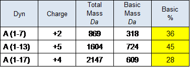
This ability is related to the charge of dynorphins that, due to the Lys and Arg residues, are the most basic naturally occurring peptides in the body (Table 1).

The remarkable role played by the Lys-Arg basic pair is yet assessable in the first Pro-Dyn demolition, resulting in Dyn A (1-17) and Dyn B (20-32) release. The cleavage is exerted by the selective activity of the Proprotein Convertases (e.g. PC2), a family of enzymes that belong to the Subtilisin/Kexin-type family, with activity at dibasic C-terminal sequence summarized as:
Lys-Arg-|-xaa > Arg-Arg-|-xaa (xaa = generic amino acid)
A Kexin-like enzyme (e.g. Kex2) could be also involved in the second Dynorphin cleavage, providing – despite the previous “in vitro” studies (Yu, 1996) – an “in vivo” fragment such as the Dyn A (1-7) (Reed, 2003), herein hypothesized as the endogenous vector of the promising prodrug conjugated with quercetin.
This flavonoid should represent one of the two relevant bioactive elements, having it a proven therapeutic activity when used both in cancer prevention and treatment.
Highly ubiquitous and well-classified dietary polyphenol found in various fruits and vegetables, quercetin (3,3′,4′,5,7-penta-hydroxyflavone) and its various metabolites have been widely demonstrated to possess an antiproliferative and proapoptotic activity in various cancer cell types (Harris, 2016).
Taking into account what has been reported in the literature on the endogenous opioid peptides (e.g. Dyn A (1-7)) and the dietary polyphenols (e.g. quercetin), this study proposes a possible combination of the two molecules into a novel prodrug, conjugated through a carbamate linker.
Materials / Methods
Dynorphin
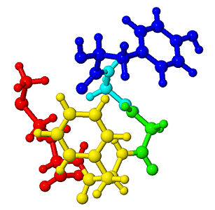
Dyn A (1-7) 3D model having a mass of 869 Da has been initially derived from the Met-Enkephalin structure shown in Figure 1 (Mouret, 2012), by modification at C-terminal site with a Met to Leu substitution. For the current study and starting from the more recent Dyn A (1-13) conformation in Figure 2 (O’Connor, 2015), the derivative N-terminal YGGFLRR structure representing the Dyn A (1-7) has been used, after deletion of the C-terminal fragment IRPKLK.
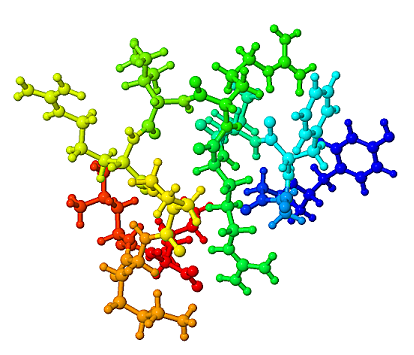
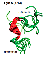
In detail, the reference structure of the dynorphin A (1–13) peptide (YGGFLRRIRPKLK) bound to the human kappa opioid receptor (KOR) has been determined by liquid-state NMR spectroscopy (O’Connor, 2015). Interestingly, the N-terminal opioid Y1-G2-G3-F4 was resulted to be flexibly disordered. The central part of the peptide L5-R6-R7-I8-R9, which forms a helical turn, was observed to be stable. The C-terminal segment (P10-K11-L12-K13), like the N-terminal, was still observed flexibly disordered (Figure 3).
Due to the possible influence of the peptides basic charge in the interaction on cell membrane (Marinova, 2005), the most interesting Dyn fragments including Dyn A (1-7) have been also compared (Table 2) in terms of percentage of basic mass/charge component resulting on them.
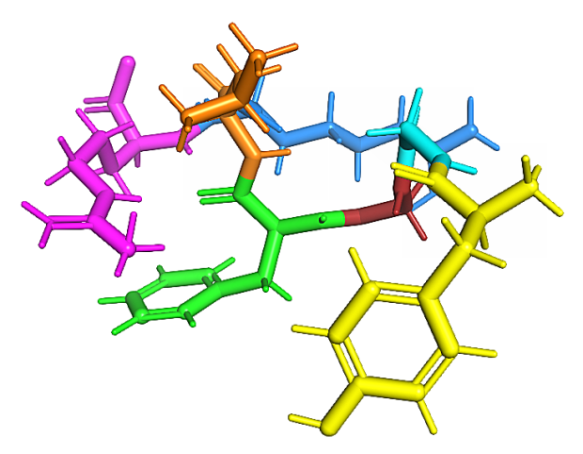
According to the proposed approach, the study on the endogenous component involved in prodrug, as derived from the Dyn A (1-13), is still focused on Dyn A (1-7) fragment (Figure 4).
Quercetin
Quercetin is a polyphenol belonging to the flavonol subgroup of flavonoids. Widely distributed in plants, vegetables, and fruits, it exhibits a variety of therapeutic properties, such as antioxidant, radical-scavenging, anti-inflammatory, antibacterial, antiviral and immunomodulatory (Yao, 2016).
Besides, quercetin can prevent neurological disorders and exerts protection against mitochondrial damages. (Massi, 2017). Numerous in vitro studies as an anti-cancer agent (Harris, 2016) via tyrosine kinases inhibition (Kim, 2009), (Russo, 2014) and as a suitable candidate in cell cycle control and of tumor therapy (Nam, 2016) (Poór, 2016) have been also available.
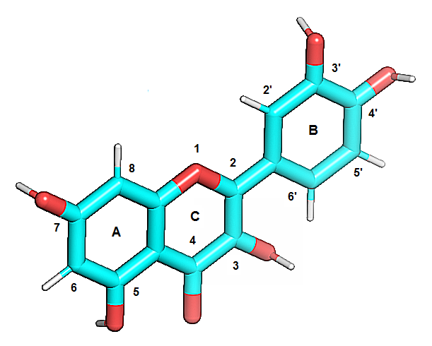
The bioavailability of quercetin in humans is low, highly variable, and rapidly cleared with an average terminal half-life of 3.5h (Li, 2016) after ingesting dietary food, drinks, or supplements. Due to its lipophilic nature, quercetin is also able to pass with ease through cell membranes. Its structure consists of two aromatic rings (A and B), linked through a pyranic ring (C) with five hydroxyl groups (–OH) attached to 3, 5, 7, 3’ and 4’ positions (Figure 5).
In plants, quercetin can exist in either free (aglycone) or bounded form, mainly with carbohydrates (quercetin glycosides) and alcohols, mostly methanol (quercetin methyl ethers), while less frequently occurring are quercetin derivatives featuring prenyl and sulfate substituents (Massi, 2017).
Carbamate
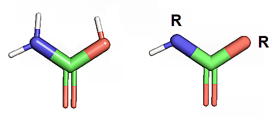
The carbamate group (Figure 6) plays an important role in modern drug discovery. Thanks to their chemical stability and ability to permeate cell membranes, carbamates (or urethanes) are widely utilized as peptide bond surrogates in medicinal chemistry applications, in many therapeutic agents, and in carbamate-based anticancer drugs (Ghosh, 2015).
The use of a carbamate as a convenient linker in prodrugs based on flavonoids (Resveratrol) coupled with amino acids has been recently proposed (Mattarei, 2015). All the synthesized prodrugs, in which the hydroxyl groups of Resveratrol were engaged in an N-monosubstituted carbamate linkage with Leu, Ile, Phe, and Thr residues, have been also characterized showing a good in vivo solubility and stability in aqueous media and in blood.
Moreover, a rational design and structure-activity relationship studies of four quercetin–amino acid hybrids (Que-Ala, Que-Leu, Que-Glu, and Que-Phe) targeting the anti-apoptotic protein Bcl-xL has been also performed very recently (Kellici, 2017). In this hypothesis and through a carbamate bond, all conjugates were made by direct coupling of the α-amine of the amino acids to the phenol groups located in ring B of quercetin.
Discussion
Even if detected in rats by Reed et al. in 2003 but not investigated further, Dyn A (1-7) may represent an active peptide able to interact with cell membranes, via a non-opioid mechanism, or through the lipid bilayers, by transients pores formation. Due to its basic characteristics and low molecular mass (869 Da), YGGFLRR administration is supposed to exert a tonic effect on cell cycle regulation and mutagenesis, when a poor amount of dynorphins occurs in the human body.
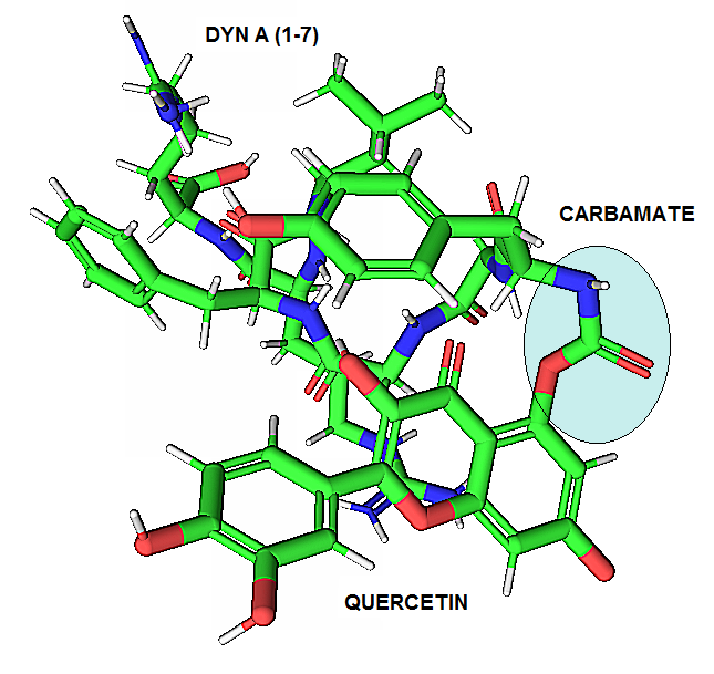
As suggested in the previous study, Dyn A (1-7) may have a beneficial role not only alone but also when combined with dietary polyphenols (e.g. quercetin). In this scenario and in order to propose a possible complex, the recent role offered by carbamates and carbamate-derived compounds in medicinal chemistry and their widespread employment as drugs and prodrugs have been evaluated as a (Tyr1) Dyn-Que conjugate through an R-NH(CO)O-R bond.
The resulting 3D stick conformation of the Dyn A (1-7)–quercetin complex has been modeled with PyMOL (DeLano, 2002) and proposed in Figure 7.
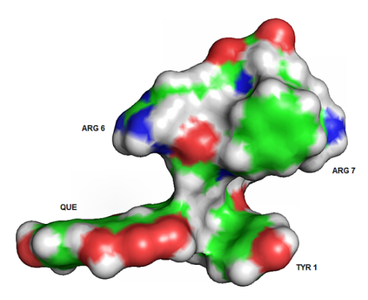
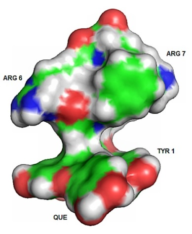
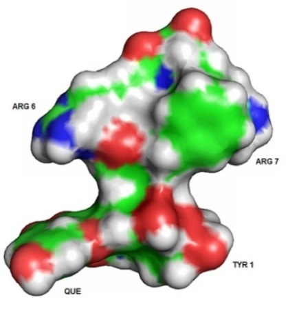
The surface conformation of Dyn-Que complexes, when bounded at the 3-OH, 5-OH, and 7-OH of quercetin, have been also evaluated and shown in Figures 8a, 8b, and 8c respectively.
Summary
Generally addressed to the Dyn-Que conjugate, the present study along with the previous ones [1] [3], proposes the presence of this compound in the human body, either as naturally occurring (i) or as prodrug conjugate (ii).
(i) Endogenous – Although their presence in human tissues is currently undetected, the complex formation may be imputable to the interaction between the endogenous opioid peptide YGGFLRR dynorphin A (1-7) and quercetin (or similar), a natural product of the flavonoids family.
(ii) Prodrug – Thanks to carbamate bridge, the conjugate dynorphin A (1-7)-quercetin may be also supposed as a prodrug, beneficially active in cell-cycle regulation or against cell dysregulation and cancer when a poor amount or lack of Dyn-Que occurs.
I hope that this study will stimulate further investigation on the peptide-carbamate-flavonoid motif, in a perspective of a new medicinal chemistry approach toward novel prodrugs and related initiatives against human cancer.
Records
[1] Report: “Methadonine: a new compound for cancer prevention and cure”. Bosica R. et al. (Jun 2002).
[2] Website (italian only): http://xoomer.virgilio.it/boroinfo.net Issue/Rev. (Jan 2004); (Jun 2004); (Aug 2005). NO LONGER AVAILABLE
[3] Report: “Methadonine – Facts and hypotheses regarding a new endogenous molecule involved in human cancer”. Bosica R.(Jan 2012).
[4] Website (Italian/English): http://www.webalice.it/roberto.bosica Issue/Rev. (2012). NO LONGER AVAILABLE
[5] Report (current work): “Hypothesis on dynorphin-quercetin conjugates as novel prodrugs against human cancer”. Bosica R. (Feb. 2018).
References
DeLano, W. L. (2002). PyMOL Molecular Graphics System. DeLano Scientific, San Carlos, CA, 700.
Fazalul Rahiman S S, Morgan M, Gray P, Shaw P N and Cabot P J. Dynorphin 1-17 and its n-terminal biotransformation fragments modulate lipopolysaccharide-stimulated nuclear factor-kappa b nuclear translocation, interleukin-1beta and tumor necrosis factor-alpha in differentiated thp-1 cells. PLoS ONE (2016), 11(4): e0153005.
Ghosh A K and Brindisi M. Organic carbamates in drug design and medicinal chemistry. J. Med. Chem. (2015), 58, 2895−2940.
Harris Z, Donovan M G, Branco G M, Limesand K H and Burd R. Quercetin as an emerging anti-melanoma agent: a four-focus area therapeutic development strategy. Frontiers in Nutrition (2016), 3:48.
Kellici T F, Chatziathanasiadou M V, Lee,M-S, Sayyad N, Geromichalou E G, Vrettos E I, Tsiailanis A D, Chi S-W, Geromichalos G D, Mavromoustakos T and Tzakos A G. Rational design and structure–activity relationship studies of quercetin–amino acid hybrids targeting the anti-apoptotic protein. Bcl-xL Org. Biomol. Chem. (2017), 15, 7956.
Kim M K, Park K, Yeo W, Choo H and Chong Y. In vitro solubility, stability and permeability of novel quercetin–amino acid conjugates. Bioorganic & Medicinal Chemistry 17 (2009), 1164–1171.
Li Y, Yao J, Han C, Yang J, Maria Chaudhry M T, Wang S, Liu H and Yin Y. Quercetin, Inflammation and Immunity. Nutrients (2016), 8, 167.
Marinova Z, Vukojevic V, Surckeva S, Yakovleva T, Cebers G, Pasikova N, Usynin I, Hugonin L, Fang W, Hallberg M, Hirschberg D, Bergman T, Langel U, Hauser KF, Pramanik A, Aldrich JV, Graslund A, Terenius L and Bakalkin G. Translocation of dynorphin neuropeptides across the plasma membrane: a putative mechanism of signal transmission. JBC Paper in Press (2005), 280:28, 26360–26370.
Massi A, Bortolini O, Ragno D, Bernardi T, Sacchetti G, Tacchini M and De Risi C. Research progress in the modification of quercetin leading to anticancer. agents Molecules (2017), 22,1270.
Mattarei A, Azzolini M, La Spina M, Zoratti M, Paradisi C and Biasutto L. Amino acid carbamates as prodrugs of resveratrol. Scientific Reports (2015), 5:15216.
Maximyuk O, Khmyz V, Lindskog C J, VukojevićV, Ivanova T, Bazov I, Hauser K F, Bakalkin G and Krishtal O. Plasma membrane poration by opioid neuropeptides: a possible mechanism of pathological signal transduction. Cell Death and Disease (2015), 6, e1683.
Mouret L. 2LWC – Met-enkephalin in DPMC SUV”. RCSB / PDB (2012). http://www.rcsb.org/pdb/explore.do?structureId=2lwc
Nam J S, Sharma A R, Nguyen L T, Chakraborty C, Sharma G, and Lee S S. Application of bioactive quercetin in oncotherapy: from nutrition to nanomedicine. Molecules (2016), 21,108.
O’Connor C, White K L, Doncescu N, Didenko T, Roth B L, Czaplicki G, Stevens R C, Wüthrich K and Milon A. NMR structure and dynamics of the agonist dynorphin peptide bound to the human kappa opioid receptor. PNAS (2015), 112:38, 11852–11857.
RCSB / PDB (2015) http://www.rcsb.org/pdb/explore.do?àstructureId=2n2f
Poór M, Zrinyi Z and Kőszegi T. Structure related effects of flavonoid aglycones on cell cycle progression of HepG2 cells: Metabolic activation of fisetin and quercetin by catechol-O-methyltransferase. (COMT) Biomedicine & Pharmacotherapy (2016), 83, 998-1005.
Reed R, Zhang Y, Chait B T and Kreek M J. Dynorphin A(1–17) biotransformation in striatum of freely moving rats using microdialysis and matrix-assisted laser desorption/ionization mass spectrometry. Journal of Neurochemistry (2003), 86, 815–823.
Russo G L, Russo M, Spagnuolo C, Tedesco I, Bilotto S, Iannitti R, Palumbo R. Quercetin: a pleiotropic kinase inhibitor against cancer. Cancer Treat Res. (2014),159:185-205.
Sankararamakrishnan R and Weinstein H. Structure-based differences in the membrane insertion properties of Dynorphin A(1-13) and Des-Tyr dynorphin. Poster of Mount Sinai School of Medicine, New York 10029 and Biophysical Journal (2001), 543A-544A.
Uezono T, Toraya S, Obata M, Nishimura K, Tuzi S, Saitô H and Naito A. Structure and orientation of dynorphin bound to lipid bilayers by 13C solid-state NMR. Journal of Molecular Structure (2005), 749:13-19.
Yu J, Butelman E R, Woods J H, Chait B and Kreek M J. In vitro biotransformation of Dynorphin A (1-17) is similar in human and rhesus monkey blood as studied by Matrix-Assisted Laser Desorption / lonization mass spectrometry. JPET (1996), 279:507-514.