METHADONINE: FACTS AND HYPOTHESES REGARDING A NEW ENDOGENOUS MOLECULE INVOLVED IN HUMAN CANCER – (LA METADONINA: FATTI ED IPOTESI RIGUARDANTI UNA NUOVA MOLECOLA ENDOGENA COINVOLTA NEI TUMORI UMANI)
Record of Changes
Initial Work: “Methadonine: a new compound for cancer prevention and cure”, June 2002
Website (Italian only): http://xoomer.virgilio.it/boroinfo.net – Issue/Rev. Jan 2004; Jun 2004; Aug 2005 – NO LONGER AVAILABLE
Website (Italian/English): http://www.webalice.it/roberto.bosica – Issue/Rev. 2012 – NO LONGER AVAILABLE
CONTENTS
1 – INTRODUCTION
2 – MOLECULAR COMPLEX
3 – OPIOID CONTENT
4 – BIODEGRADATION OF DYNORPHIN PRECURSOR
5 – DYNORPHIN A (1-7)
6 – DYNORPHIN FRAGMENT CHARACTERISTICS
7 – RECEPTORS AFFINITY
8 – OPIOID PEPTIDE AND DISEASES
9 – OPIOID GROWTH FACTOR
10 – FLAVONOID STRUCTURE
11 – FLAVONOID PROPERTIES
12 – FLAVONOIDS IN THE HUMAN BODY
13 – GENETIC INVOLVEMENT
• Chromosome 7 and CFTR protein
• Influences on the pancreas
• Chromosomal fragile sites
• Chromosome 17
14 – DISCUSSION
15 – PERSPECTIVES
16 – ACKNOWLEDGEMENTS
17 – REFERENCES
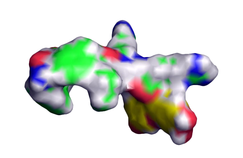
1. INTRODUCTION – The purpose of this study is to explore the formation and the role played by Methadonine and its two components in humans, through the experience shown by a case report of endometrial cysts, nasal polyps, and breast cancer in patients with possible genetic disorders and related neuroendocrine deficiencies. Methadonine is the name assigned to a new molecule still undetected in the human body which affects the cell cycle regulation and the possible mechanism of cell mutations and cancer when a lower amount or lack of the molecular elements occurs. Its formation (Figure 1) is mainly due to the interaction between the endogenous opioid peptide Dynorphin A(1-7) and Quercetin, a naturally occurring compound belonging to the Flavonoid family.
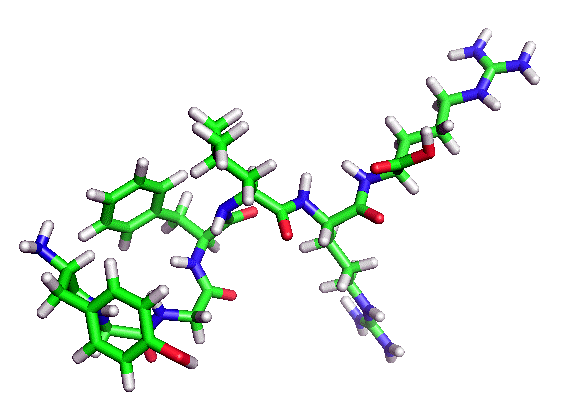
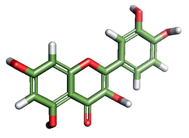
2. MOLECULAR COMPLEX – On the peptide, and presumably promoted by an enzymatic activity ATP-dependent, a covalent bonding of the Flavonoid with Tyrosine (Tyr1) or less probably with Phenylalanine (Phe4) should preferably take place.
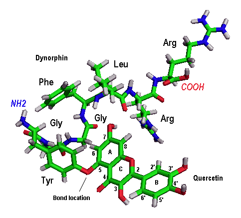
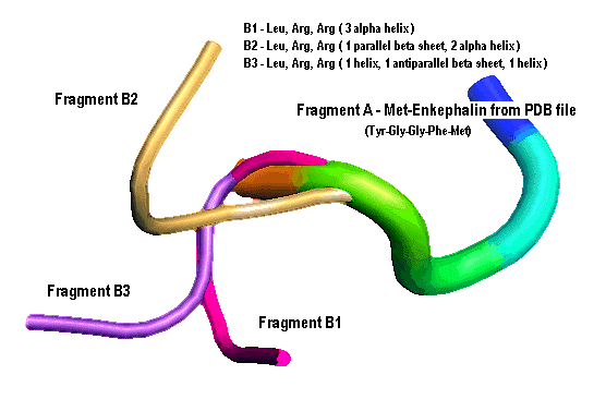
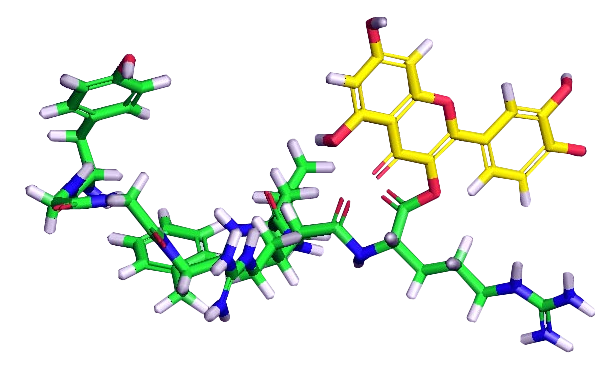
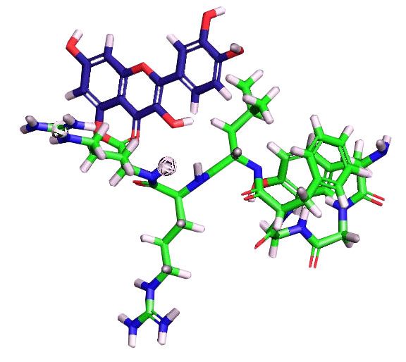
In the event of bonding with the Tyrosine (Figure 2), we should observe the formation of a molecular bridge (and H2O release) based on a single oxygen atom, resulting from the activity of the hydroxyl group of Tyrosine (phenolic OH) and the OH group (position 5) of Quercetin. By using the online B (Biomer) and PyMOL programs, a conformational 3D structure of the pepdite was built by similarity to the Dynorphin and relevant initial Met-Enkephalin conformation (Marcotte, 2004). Therefore, in addition to the 1-4 Tyr-Gly-Gly-Phe Enkephalin peptide presented in Fragment A (Methionine not considered), the structural model (Figure 3) was then sequenced with the 5-7 remaining Leu-Arg-Arg residues in Fragment B (stick model B3 selected).
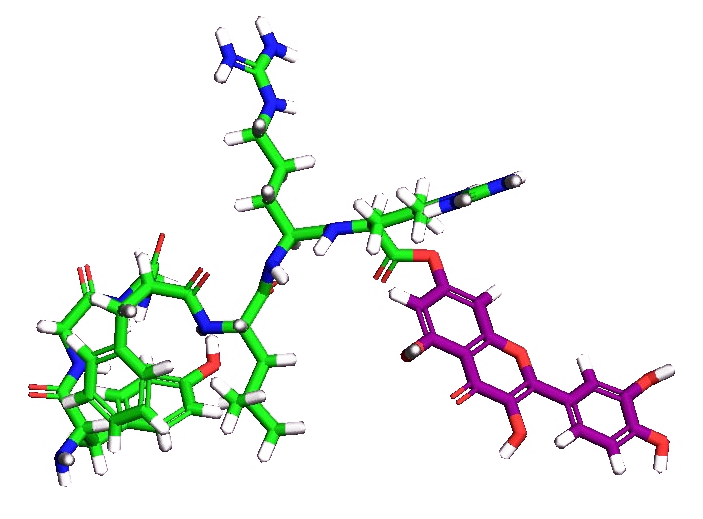
The molecular setup of the side chain was completed with the integration of the planar Quercetin (Kim H, 2006), formed in plants by biotransfomation of both Tyrosine and Phenylalanine precursors (Middleton, 2000). Besides the hypothesis put forward for the Dynorphin-Quercetin complex formation at Tyr1 level, Quercetin bonding at peptide C terminal (Arg7) has been also evaluated (Figures 4). This approach is based on prodrug studies regarding the ability of Quercetin to interact with amino acids at the 3-OH or 7-OH group (Kim MK, 2009) and to increase its pharmacokinetic properties such as water solubility, stability, and relevant prodrug potential administration.
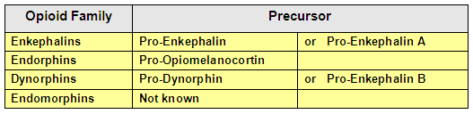
3. OPIOID COMPONENT – Dynorphins, together with Enkephalins and Endorphins (Table 1), are neurotransmitters belonging to the family of endogenous opioid peptides (Koneru, 2009) synthesized in the human CNS. The Dynorphin active peptides, such as Fragment A(1-7), are parts of Pro-Dynorphin (Tables 2), a precursor of 254 amino acids primarily present in the hypothalamus.
As recently observed, Dynorphins have the ability to penetrate the plasma membrane (Marinova, 2005) by stable or transient pores, similar to what has been demonstrated for CPPs (Cell-Penetrating Peptides). Dynorphins have a structural similarity to CPPs, in possessing a high content of basic amino acids and especially a large number of Arginine residues. So that, together with Dyn A(1-7), they are probably the most basic neuropeptides to our knowledge (Marinova, 2006).

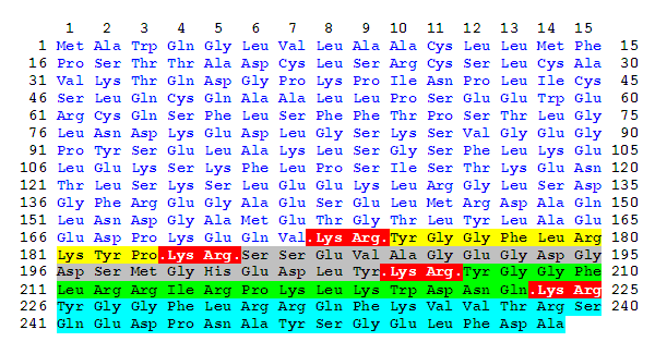
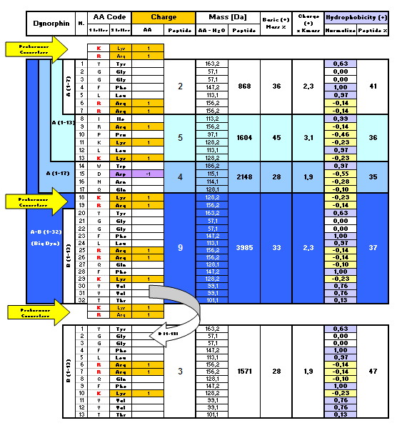
4. BIODEGRADATION OF DYNORPHIN PRECURSOR – The initial Pro-Dynorphin degradation, supposed at pancreatic level, is probably obtained by a Trypsin-like enzymatic activity which recognizes, with different intensity, the pair of amino acids Lys-Arg (or KR) basic residues (Tables 2). The preferential cleavage sites should be sequenced as follows:
Lys-Arg-|-xaa > Arg-Arg-|-xaa > Lys-|-xaa > Arg-|-xaa
(Note: xaa = generic amino acid)
The hypothesis that the pancreas may represent a site for Pro-Dynorphin demolition, and/or a local source of enzymes for protein processing, is supported by the fact that both Dynorphin, isolated in the pancreatic cells (Steele, 1989; Cetin, 1990), and the enzyme calcium-dependent Pro-Hormone Convertase 2 (PC2) (Ugleholdt, 2004), are both localized into the secretory granules of Pancreas.
PC2 belongs to one of the seven protein convertases subtypes, an enzymatic family that plays a crucial role in various physiological processes related to several diseases, among which cancer (Taylor, 2003). It also operates selectively in various precursors biotransformations (Pro-Dynorphin included), when the preferential pair of Lys-Arg-|-xaa amino acids are recognized. Based on the selective Lys-Arg cleavage sites shown above, the list of peptides delivered from the first Pro-Dynorphin demolition is reported in Table 3, in terms of numerical and amino acids sequence identifications.
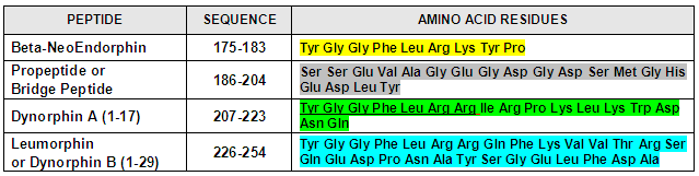
5. DYNORPHIN A (1-7) – With regard to the subsequent processing of Dynorphin A(1-17) linked to the fragment 207-223 (Table 3), a specific Proteinase was firstly isolated from the human spinal cord (Silberring, 1992) and subjected to exclusive conversion to Leu-Enkephalin and to the relevant COOH-terminal extensions Leu-Enkephalin-Arg6 (which was a major conversion product) and Leu-Enkephalin-Arg6-Arg7 (Table 4). More recently, a further study for investigating the biotransformation of Dynorphin A(1–17) in the rat Striatum in vivo (Reed, 2003) was performed. Through the direct infusion of this peptide in rats freely moving, followed by MALDI (Matrix-Assisted Laser Desorption / Ionization) mass spectrometry identification, the formation of two complementary peptides Dyn A (1-7) and Dyn A (8-17) has been observed.
The enzyme responsible for these fragments formation, different from those so far studied as Protein Convertase 2, Carbossilpeptidase E, Dynorphin A Converting Enzyme, and Dynorphin A(1-17) Processing Enzyme, remains to be determined. Although not yet detected in humans, the enzymatic activity capable of forming Dynorphin A(1-7) could be imputable, as a single and highly selective trypsin-like process, by the calcium-dependent membrane protein Kexin (Kex2) Protease, active in fungi/yeast (Jalving, 2000; Vilaça, 2008), at intracellular level (Golgi apparatus) and able to cleave the C-terminal dibasic Lys-Arg-|-xaa and Arg-Arg-|-xaa amino acid sites.
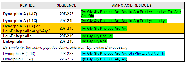
6. DYNORPHIN FRAGMENTS CHARACTERISTICS – Compared to their low molecular masses, Dynorphin fragments are characterized by a high content of basic and hydrophobic (core) residues. Apart from Dyn B(1-13), all the peptides considered (Table 5) are probably capable of penetrating the cell through transient pores (Marinova, 2005 and 2006; Ugonin, 2007) or ion channels, reaching a rapid penetration in 1-5 minutes after in vitro incubation. Therefore, the ability of Dyn A peptides to translocate across plasma membrane seems to be linked to the content (about 35%) of basic and hydrophobic amino acids for whom the fragments are characterized.
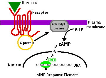
7. RECEPTORS AFFINITY – Opioid peptides act selectively as agonists or antagonists toward the relevant seven-domain G-protein coupled receptors located in the plasma membrane of cells (Figure 5). The prerequisite for the Opioid Receptors activation is the selective binding of peptides possessing, as a typical motif, the four N-terminal amino acids residues Tyr1-Gly2-Gly3-Phe4. In addition to the role exerted by Tyr1and Phe4, the residual sequences associated with Enkephalins, Endorphins, or Dynorphins, are responsible for their selectivity towards the different types of opioid receptors defined as delta, mu, and kappa receptors.
Kappa Opioid Receptors (KORs) are particularly sensitive to the pair of Arg6-Arg7 basic residues, or, more generally, to the first seven amino acids of the Dynorphin A (1-17) peptide (Jordan, 2000). KORs are widely distributed in the body, since their presence has been demonstrated, not only at Central Nervous System (CNS) level, but also in the pancreas, liver, digestive tract, endometrium, and mammary glands (Khawaja, 1990), (Chatzaki, 2001). In addition to the receptor affinity expressed by the seven N-terminal residues YGGFLRR, Dynorphins assume particular relevance for their ability in Protein Kinase control through a phosphorylation process, and for the activation of KORs signaling internally the cell without any receptor internalization (Jordan, 2000).
8. OPIOID PEPTIDES AND DISEASES – The presence of opioid peptides and their precursors was studied in 108 different types of neuroendocrine and non-neuroendocrine tumors (Bostwick, 1987). By using specific antibodies, it was possible to observe a consistent cytoplasmic immunoreactivity of the opioid tetrapeptide Tyr-Gly-Gly-Phe in several cell lines of carcinoids tumors (18 cases of 26), variously located in the pancreatic islets (7 of 10), parathyroid adenomas (8 of 9), etc. On the basis of the immunoreactive response observed in non-neoplastic cells (i.e pancreatic islets, pituitary, intestinal plexus, etc.), it was also assumed that the increased expression of opioid peptides in malignant cells may be related to the pathology of cancer.
Further confirmation about the relationship between endogenous opioids and cancer comes from the study on other 63 cell lines of benign and malignant breast cancer (Chatikhine, 1994). Among the infiltrating ductal carcinomas analyzed in the study, 93% of them were found positive for the presence of Beta-Endorphin, 87% for Leu-Enkephalin, and 90% for the peptide Met-Enkephalin.
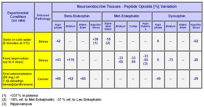
Moreover, some in vivo studies performed on laboratory animals confirmed a strong variation of physiological levels of Beta-Endorphin, Met-Enkephalin, and Dynorphin (Table 6) in individuals subjected to induced stressful or pathological events (Vaswani, Jan 1986; May 1986; 1988). About the data related to neuroendocrine tissues shown in Table 6, it is interesting to note a potential dual body’s response to stressful events, with a general increase of Beta-Endorphin corresponding to an evident reduction of Leu-/ Met-Enkephalin and Dynorphin. Moreover, plasma levels of Met-Enkephalin were found 3.5- to 7.9-fold less in transplanted mice with pancreatic and colon cancer cells than in control mice (Zagon, 2006).
As for the Dynorphins in the human body, significantly lower values of these peptides were also found in the menstrual blood of patients affected by endometriosis (Yu, 1993), a pathological status that implies the presence of endometrium (the mucous lining the uterus) in improper extrauterine locations such as ovaries, fallopian tubes, and intestines. Because of the possible infiltration of mucus in the endometrial cells and subsequent migration through the blood and lymphatic vessels, relevant forms of inflammatory injury and mucosal thickening can occur, with a difficult diagnostic prediction, to lungs or nasal polyposis. Endometriosis is also associated with the ability to develop, in addition to cysts and cancer localized in the primitive sites, several forms of peripheral cancer like melanoma and breast cancer (Swiersz, 2002).
The polycystic ovarian syndrome, a neuroendocrine defect observed in clinical reproductive disorders, is still associated with Dynorphin peptides localized, as well as Kisspeptin and Neurokinin B, in the arcuate nucleus cells of the hypothalamus (Lehman, 2010). The relationship between the expression-activity of the opioid peptides and their possible impact on human diseases have been examined in order to support the case report here studied.
9. OPIOID GROWTH FACTOR – Because of their structural similarity, a strong correlation between Dynorphin fragments (Table 4) and the cognate Opioid Growth Factor (OGF), synonymous with Met-Enkephalin (YGGFM), also exist. The endogenous OGF, the native opioid peptide mainly derived from the Pro-Enkephalin precursor, serves as the active negative growth factor in cancer diseases (Zagon, 2006 and Cheng, 2007). It affects, according to the relevant OGF-OGF receptor axis, the cell cycle regulation (G0-G1 phase), and the control of cell proliferation through the Kinases and the inhibitory DNA synthesis pathway (Martin-Kleiner, 2004).
This evidence is confirmed by the presence of the mentioned receptors (OGFr), both at the free cytoplasm level and on the outer surface of the cell nucleus (Swiss-Prot: OGFR_HUMAN, Q9NZT2, and related links). The role expressed by Dynorphins as regulatory peptide hormones has been also demonstrated by fragment A (1-8). When placed with the serum-free culture of mouse neuroblastoma cells, the sequence YGGFLRRI produced beneficial effects on cell cycle regulation of DNA and total cellular protein in the direction of delaying neuronal aging (Sun, 1995).
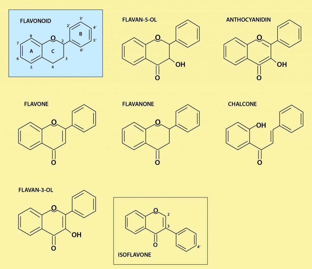
10. FLAVONOID STRUCTURE – Flavonoids are a large group of natural substances with low molecular weight, widely distributed in nature in more than 4000 structural variants (Middleton, 2000). Also recognized as Polyphenols, they are responsible for autumnal pigmentation of plants and yellow, orange, and red colors in flowers, fruits, and vegetables. In plants, Flavonoids are synthesized from the aromatic amino acids Phenylalanine and Tyrosine (Middleton, 2000). After modification, due to light- and temperature-dependent process, the final structure (Figure 6; Figure 7) is characterized by a typical C15 core, built on two benzene (A) and (B) rings connected, through a double bond, to an intermediate (C) ring resulting either as Pyran (i.e. anthocyanins) or gamma-Pyrone (i.e. flavones).
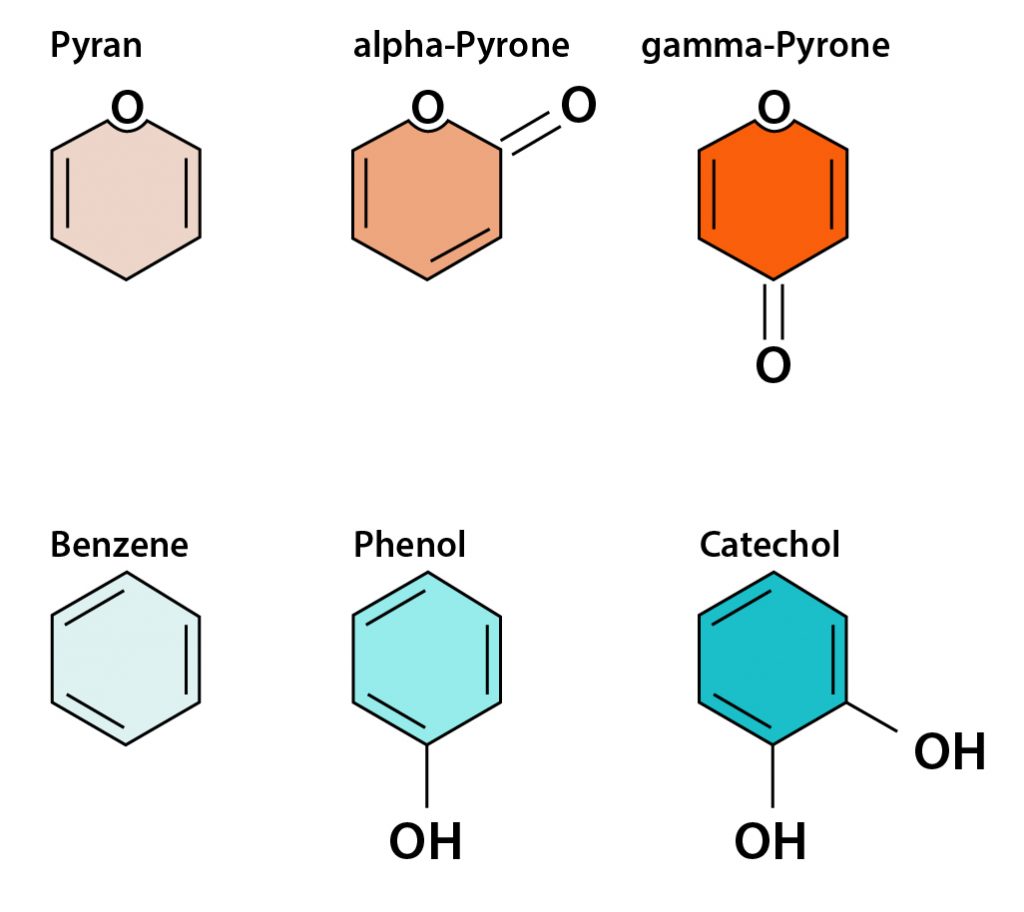
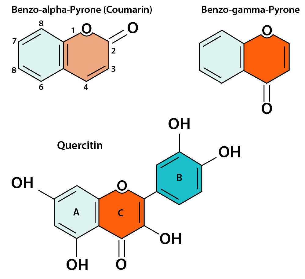
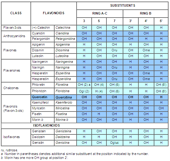
The isomer alpha-Pyrone, instead, occurs in Coumarins (Figure 6), a group of phenolic compounds where the pair of (A) (C) planar rings is called Benzo-alpha-Pyrone. Both Coumarins and Coumarin-related compounds (Flavonoids), of which these rings are constituents, belong to the family of Benzopyrones and/or Polyphenols (Figure 7, Table 7), widely included in dietary exposure of foods and beverages.
11. FLAVONOID PROPERTIES – Thanks to the high reactivity of the several hydroxyl groups (OH), the flavonoids are known as antioxidant substances capable of inactivating free radicals. The antioxidant activity is exerted on the oxygen-reactive molecules that may cause DNA damages, abnormal cell division, and cancer (Nijveldt, 2001). The interaction with the DNA has been also confirmed by using the basic structure of Flavones, through the levels of fluorescence and absorption which result from the direct binding to DNA (Dean, 2003). Assuming R’ as the free radical and O’ as the oxygen free radical related to the oxidized Flavonoid, these compounds exert their function under the following condition:
Flavonoid (OH) + R’ > Flavonoid (O’) + RH
Besides the extensive studies performed to prove the beneficial antioxidant, anti-inflammatory, antiviral, immunomodulatory, and anticancer properties (Lacy, 2004; Ong, 2004; Zhou, 2010), Flavonoids have been also investigated for their ability to exhibit a wide range of properties regarding the fluorescence emissions.
In Coumarins, the intrinsic fluorescence is extremely sensitive to the local environment of the molecule (Wagner, 2009), whereas in Flavones (i.e. Quercetin) this property allows the identification of cellular target proteins, offering the opportunity to study and predict the biological and pharmacological effects in humans (Gutzeit, 2005; Nifli, 2007). In addition, the well-known Protein Tyrosine Kinase (PTK) inhibitory role (at micromolar level) of Quercetin (Huang, 2009) and its ability for binding Human Serum Albumin (HAS) (Rolinski, 2007) have been both demonstrated.
12. FLAVONOIDS IN THE HUMAN BODY – Flavonoids (Table 7) are usually assumed in diet, mostly in the “Mediterranean Diet” which is particularly rich in olive oil, citrus, and vegetables. The values ingested, mainly referred to as Flavonols and Flavones glucoside (with sugar molecule), are strongly influenced by the food habits adopted in each country. Despite the average flavonoids intake observed in the past as 23 mg/day (Hertog, 1993), the lowest being in Finland (2.6 mg/day) and the highest (Nijveldt, 2001) in Japan (68.2 mg/day), recent studies widely revised previous data.
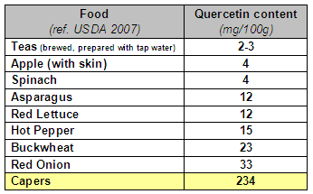
Based on the expanded number of polyphenols included now in the analysis, the revised data imply actually that the average amount of Favonoids in diet are higher (Chun, 2007; Zamora-Ros, 2010), and estimated at about 250 mg/day (i.e. 209 in Finland, 243 in US and 313 in Spain). Among the Flavonoids assumed in diet (USDA, 2007), the flavonol Quercetin is the most abundant and present especially in foods listed in Table 8. It is interesting to note that the raw capers contain a very high amount both of Quercetin (234 mg/100g) and Kaempferol (259 mg/100g).
Although widely studied in recent decades, the role of flavonoids in humans has to be better clarified. In particular, some answers are expected on how the Flavonoids could enter the cells and whether they could accumulate in certain cell tissues (Middleton, 2000), what is the nature of the chemical interaction resulting from the presence of Quercetin in the blood (Day, 2001) and what is the molecular basis able to generate the beneficial effects of Flavonoids (Dean, 2003).
With regard to Quercetin in the blood, the red blood cells have been supposed as sites of natural accumulation and rapid storage, through an initial mechanism of passive diffusion then followed by direct bonding to hemoglobin (Fiorani, 2003). This supports the fact that red blood cells can transport Flavonoids through the bloodstream to all tissues. As for the plasma, the concentration of Flavonoids in the medium is still debated and taken at the time in about 1 micro-Mole, among populations with large consumption of fruits and vegetables (Kuo, 2002).
This circulatory level, relatively low in comparison with the dosages observed in human trials, is justified by the fact that Flavonoids may be first transmitted and then temporarily stored within the cells which operate as sites of accumulation. To complete the scenario that supposes the formation of Methadonine through the biological affinity of its two constituents, recent studies have shown that some peptide hormones, including Met-Enkephalin and Leu-Enkephalin, exert antioxidant activities similar to the flavonoids, by means the involvement of the aromatic residues as Tryptophan, Phenylalanine, and Tyrosine in particular (Moosmann, 2002; Zhao, 2004).
13. GENETIC INVOLVEMENT – This case report proposes the hypothesis regarding the modulatory role exerted by Methadonine and its constituents in humans, and describes a possible correlative pathway for a physiopathological scenario sequencing endometrial cysts, nasal polyps, and breast cancer occurrences in patients affected by neuroendocrine and genetic disorders. The genetic involvement here considered is mainly related to Chromosomes 7 and 17.
Chromosome 7 and CFTR protein – Beyond the antioxidant properties of polyphenols already discussed in this work, it is important to remember that some flavonols (such as Apigenin, Quercetin, Kaempferol) and isoflavones (e.g. Genistein, Daidzein) were also studied for their biological activities on the cell membrane, and in particular, for the ability in activating the protein Cystic Fibrosis Transmembrane Conductance Regulator (CFTR) (Schultz, 1999).
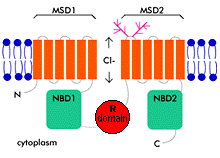
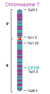
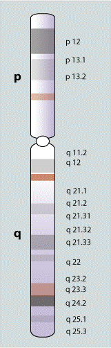
This glycoprotein (Figure 8), an ATP-binding cassette (ABC) transporter, which consists of 1480 amino acids and a molecular weight of 168 KDa (unglycosylated), is encoded by the homonym CFTR gene which has been isolated, since 1989, in the long arm (q) of chromosome 7 (Figure 9), at specific 7q31.2 In addition, CFTR is a multifunctional protein which has been given great importance, both for its influence on the bioelectric activity of the cell membrane, and for the ability to transport small molecules and peptides populate the extra- and intracellular fluid. CFTR acts as a regulator for various ion channels (pores) and the chloride channel (Cl–) in particular.
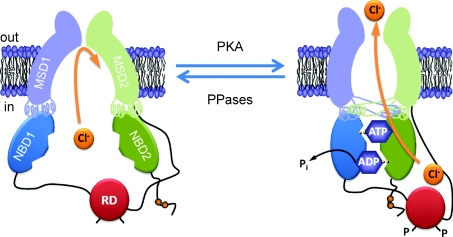
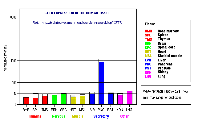
CFTR has also multi-drug resistance (MDR) features (Luckie, 2003) and influences the effectiveness of drugs and chemotherapy when administered. Similar to a biological pump, is controlled by a mechanism of phosphorylation and dephosphorylation ATP- ADP-dependent (Figure 10), carried on by enzymes (Hwang, 2009) protein kinase (open pore), and protein phosphatase (closed pore). In the body, the protein is widely distributed in tissues (Figure 11) and sites, such as red blood cells, lymph, umbilical vein, sweat glands, kidney, colon, hypothalamus, and pancreas, where CFTR reaches its maximum expression.
Extensively studied in the past for its mutations and related diseases, the CFTR gene it records to date more than 1200 different mutations. The most common mutation causing Cystic Fibrosis (CF) of the pancreas, is the ΔF508 mutation. This abnormality, an absence of three nucleotides resulting in the deletion of the Phenylalanine at position 508, is responsible for 70% of CF cases.
Another mutation identified in CFTR is G551D, which records a Glycine (G) replacing Aspartic Acid (D) in position 551 and leads the patients to the occurrence and recurrence of possible nasal polyposis. Recently, a correction in the G551D-CFTR chloride transport has been also proposed with Genistein (Zegarra-Moran, 2002). This polyphenol, belonging to the isoflavonoid family, resulted particularly potent as CFTR agonist (activator) and able to mitigate the G551D functional defect at therapeutic levels.
Influences on Pancreas – The Pancreas, one of the major constitutive elements of the neuroendocrine system, is thought to play a key role in the biodegradation of neuropeptide hormones such as Dynorphins. Moreover, most pancreatic diseases seem to be associated with genetic polymorphisms (Whitcomb, 2004). Therefore, genetic alterations that lead to disorders of the pancreas and especially in acute and chronic pancreatitis, often characterized by long latencies and uncertain symptomatic results (Transparency, 2001), should be carefully considered.
The mutations in question are mainly related to the Cationic Trypsinogen PRSS1 gene at 7q35, to the Pancreatic Trypsin Inhibitor SPINK1 gene at 5q32 and to the aforementioned gene CFTR (Salacone, 2001). Additional studies on the relationship between genetic disorders and pancreatic deficiencies (Witt, 2003) have also demonstrated that the pathological processes of this region often exhibit an altered response of the extracellular fluids, as well as the premature activation of pancreatic enzymes and mucosal thickening with the consequent obstruction of secretory ducts (Luckie, 2003).
Chromosomal fragile sites – Fragile sites are specific chromosomal regions particularly sensitive to forming constrictions, gaps, or breaks on chromosomes (Figure 12) when exposed to a specific culture and chemical conditions. They are classified as common or rare, according to their frequency in the populations. Common fragile sites are normally stable in cultured human cells. However, when cells are cultured under stressed conditions which inhibit DNA synthesis, common fragile sites reveal an impressive cluster of regions with high flexibility and low stability. On the long arm of human Chromosome 7, between 7q31 and 7q36 loci, several chromosomal aberrations have been identified over time as common fragile sites, for their induced fragility interfering with DNA replication.
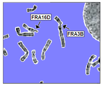
Among the 100 fragile sites estimated approximately in the human genome, the aforementioned region of the Chromosome 7 (Hellman, 2002; Glover 2005) includes in fact a remarkable number of fragile sites such as FRA7G (7q31.2), FRA7H (7q32.3), and FRA7I (7q36). In particular, the FRA7G region has been extensively studied, for the presence of genes encoding the proteins CAV1, CAV2, TAT, and CFTR, and for the relevance of the same proteins in human oncogenesis and breast cancer development (Ciullo, 2002).
Chromosome 17 – Beyond the influence exerted with Chromosome 7 alterations on cancer occurrences, some data on the aberrancies of Chromosome 17 genes (Figure 13) such as BRCA1, BRCA2, and HER2, have also been considered and associated with the physiopathological events of the present case report. On the matter, it is worth noticing that mutations, amplifications, and duplications of HER2 gene at 17q21 loci and the uncontrolled expression of the corresponding membrane protein HER2 (or C-erbB-2) in cancer lead to a worse diagnostic scenario including cancer progression and invasion.
HER2, a Tyrosine Kinase protein belonging to the Epidermal Growth Factor (EGF) receptor family, is able to control growth and cell division (Tsao, 2004). Indeed, due to the anomalous number of copies found in the corresponding gene, this receptor is frequently overexpressed in many invasive tumors (Ouyang, 1996), as well as in aggressive forms of breast cancers (Negri, 2010) characterized by an abnormal cell replication and a frequent formation of metastasis. Therefore, the detection of genomic changes of Chromosome 17 is clinically relevant not only for an accurate determination of HER2 status (Gunn, 2010) but also to difine an important prognostic marker linked to an appropriate treatment planning (e.g. Trastuzumab, thru Herceptin® and Lapatinib, by Tykerb®) against the most aggressive neoplasias
14. DISCUSSION – Supported by the scientific publications included in the reference list, this study presents a case report regarding a physiopathological scenario that includes occurrences like endometrial cysts, nasal polyps, and breast cancer (invasive ductal carcinoma), in patients with neuroendocrine deficiencies and possible genetic disorders imputable to the chromosomes 7 and 17. The hypothesis focuses on the key role played by Methadonine on human health. Although his formation and bioavailability in tissues are still undefined, the new molecule is proposed to interact in cell cycle regulation, in its possible dysregulation, and in cancer, when a lack, or a reduction of one or both molecular elements occurs. Its formation is imputable to the interaction between the endogenous opioid peptide YGGFLRR Dynorphin A(1-7) and, but not limited to, Quercetin, a natural compound belonging to the family of flavonoids.
For structural similarity, the interactions with several other flavonoids may also be possible. After the first precursor demolition performed by protein convertases (PCs), at paired of Lys-Arg sites and for releasing Dynorphin A(1-17), it is supposed that other shorter peptides, such as Dynorphin A(1-7), could be further processed. When cleaved, in the pancreas and by an Arg-Arg sensitive kexin-like enzyme (Nagamune, 1995), the active fragment A(1-7) should be then flowed rapidly into the plasma and bloodstream. Due to its intrinsic characteristics, which include a very short half-life (less than 1 minute) and a high content of basic amino acids (net charge +2) if compared to its total mass (less than 1 KDa), Dynorphin A(1-7) may be considered as a biologic missile, difficult to detect and not least, able to enter the cell via stable ion channels (e.g. chloride) or through the transient formation of pores.
With regard to the bioavailability in humans, it is believed that the level of Dyn A (1-7) may depend on the functional response of the Pancreas and that the psychopathological conditions of stress may also negatively interfere with it. In this case, the peptide could be primarily used in pain control (via Opioid Receptors), rather than in cell cycle regulation. Therefore, the deficiencies of the Pancreas and pancreatitis (see CFTR mutations), which have large latencies and difficult detections, should be considered the main causes of degenerative events such as the abnormal DNA transcription and cancer, both triggered by a low amount of Dynorphins, alone or in complex with Flavonoids.
The regulatory roles of small opioid peptides in normal cells and the apoptotic response in malignant cells have been also demonstrated through the action of Met-Enkephalin (Opioid Growth Factor, OGF) and its receptor (OGFR) axis. In other words, Methadonine represents the biological interaction in the human body between an endogenous opioid peptide (Dynorphin) and a dietary intake flavonoid (Quercetin). Both involved in the catalytic process of cells and syntheses of tissues, their bond is supposed to occur at the lipid bilayer proximity. An enzymatic involvement of a membrane protein ATP-dependant belonging to the Tyrosine Kinase family is also suspected.
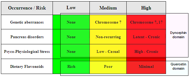
Although the questioned Dynorphin-Quercetin complex has never been detected in mammalians nor synthesized for experimental tests or for prodrug purposes, there are possibilities that Dynorphins and Flavonoids may represent together the primary elements in the extra- and intra-cellular functioning, and that their bioavailability is essential to prevent and to control cell degeneration and cancer. In order to summarize the events related to Methadonine and its physiopathological scenario described in this study, a possible occurrence/risk matrix of Table 9 has been at the end reported.
15. PERSPECTIVES – Even if widely studied in the last years, the roles of the Flavonoids and Dynorphins in humans have to be still elucidated. In particulars, some questions remain about how the Flavonoids may enter the cells and where they could accumulate in certain organs. Both the mechanism of action of the Dynorphins in cells cycle regulation and the basal amount or the possible variances of these peptides in human body have to be also deeply investigated.
Regarding Methadonine, and possible extended family of compounds based on Dynorphin A (1-7) and a generic set of flavonoids which includes Quercetin, some laboratory tests to confirm their ability in bonding or interaction should be specifically executed. In addition, the synthesis of specific prodrugs to be experiment with normal and malignant cell cultures should be similarly planned.
16. ACKNOWLEDGEMENTS – I dedicate this work to my wife Anna, to her life, and to all women who are fighting cancer. I would like to express my sincere gratitude to everyone who helped me. First of all, I would like to thank Dr. Gabriella and Dr. Alina for the time and the knowledge they have shared with me during the first phase of the pathway. And thanks, thanks, thanks to my son Francesco and my daughter Martina, the lights in the dark, always! Finally, thanks to all other… friends, so close to me.
17. REFERENCES
Bostwick DG, Null WE, Holmes D, Weber E, Barchas JD and Bensch KG. “Expression of opioid peptides in tumors”. The New England J. of Med. n. 23 (1987), 317: 1439-1443.
Cetin Y. “Immunohistochemistry of opioid peptides in the guinea pig endocrine pancreas”. Cell Tissue Res (1990), 259(2): 313-319.
Chatikhine VA, Chevrier A, Chauzy C, Duval C, d’Anjou J, Girard N and Delpech B. “Expression of opioid peptides in cells and stroma of human breast cancer and adenofibromas”. Cancer Lett. (1994), 77(1): 51-6.
Chatzaki E., Makrigiannakis A., Margioris AN, Kouimtzoglou E and Gravanis A. “The Fas/FasL apoptotic pathway is involved in k-opioid-induced apoptosis of human endometrial stromal cells”. Mol. Hum. Reprod. (2001), 7(9): 867-874.
Cheng F. “The Opioid Growth Factor (OGF)–OGF Receptor Axis Uses the p16 Pathway to Inhibit Head and Neck Cancer”. Cancer Res (2007), 67: (21).
Chun KO, Chung SJ and Song WO. ”Estimated Dietary Flavonoid Intake and Major Food Sources of U.S. Adults”. The Journal of Nutrition (2007), 137: 1244–1252.
Ciullo M, Debily MA, Rozier L, Autiero M, Billault A, Mayau V, Marhomy S, Guardiola J, Bernheim A, Coullin P, Piatier-Tonneau D and Debatisse M. “Initiation of the breakage-fusion-bridge mechanism through common fragile site activation in human breast cancer cells: the model of PIP gene duplication from a break at FRA7I”. Human Molecular Genetics (2002), 23: 2887-2894.
Day AJ and Williamson. “Biomarkers for exposure to dietary flavonoids: a review of the current evidence for identification of quercetin glycosides in plasma”. Br. J. Nutr. (2001), 86 Suppl. 1: S 105-110.
Dean J and Musich P. “Flavonoids – Keep it Simple: Mechanistic Studies of Antioxidant Activity”. The Second WCRF International Expert Report, supplement: International Research Conference on Food, Nutrition, and Cancer. J. Nutr. (2003), 133: 3851S-3868S.
Fiorani M, Accorsi A and Cantoni O. “Human red blood cells as a naturalflavonoid reservoir”. The Second WCRF International Expert Report, supplement: International Research Conference on Food, Nutrition, and Cancer. Free Radic. Res. (2003), 37(12): 1331-1338.
Glover TW, Arlt MF, Casper AM and Durkin SG. “Mechanisms of common fragile site instability”. Human Mol Genetics (2005), Vol 14 (2): R197-R205.
Gunn S, Yeh IT, Lytvak I, Tirtorahardjo B, Dzidic N, Zadeh S, Kim J, McCaskill C, Lim L, Gorre M, and Mohammed M “Clinical array-based karyotyping of breast cancer with equivocal HER2 status resolves gene copy number and reveals chromosome 17 complexity “. BMC Cancer (2010), 10: 396.
Gutzeit HO, Tokalov SV, Ludwig-Müller J and Rusak G. “Monitoring Flavonoid Metabolism in Human Cells by Exploiting Fluorescence Elicited upon Quercetin/Protein Interactions”. Croatica Chemica Acta (2005), 78 (3): 337-342.
Hellman A, Zlotorynski E, Scherer SW, Cheung J, Vincent JB, Smith DI, Trakhtenbrot L and Kerem B. “A role for common fragile site induction in amplification of human oncogenes”. Cancer Cell (2002), Vol. 1.
Hertog MGL, Hollman PCH, Katan MB and Kromhout D. “Intake of potentially anticarcinogenic flavonoids and their determinants in adults in the Netherlands”. Nutr. Cancer (1993), 20: 21-29.
Huang H, Qian J, Proffit J, Wilber K, Jenkins R and Smith DI. “FRA7G extends over a broad region: coincidence of human endogenous retroviral sequences (HERV-H) and small polydispersed circular DNAs (spcDNA) and fragile sites”. Oncogene (1998), 16(18): 2311-9.
Huang H, Jia Q, Ma J, Qin G, Chen Y, Xi Y, Lin L, Zhu W, Ding J, Jiang H and Liu H. “Discovering novel quercetin-3-O-amino acid-esters as a new class of Src tyrosine kinase inhibitors”. Eu Journal of Medicinal Chemistry (2009), 44: 1982–1988.
Hwang TC and Sheppard DN. “Gating of the CFTR Cl− channel by ATP-driven nucleotide-binding domain dimerization”. J Physiol (2009), 587.10: 2151–2161.
Initial Work:. “METHADONINE: A NEW COMPOUND FOR CANCER PREVENTION AND CURE” (June 2002).
Jalving R, van de Vondervoort PJ,Visser J, and Schaap PJ. “Characterization of the Kexin-Like Maturase of Aspergillus niger”. Appl. Envir. Microbiol. (2000), 66: 363 – 368.
Jordan BA, Cvejic S, and Devi LA. “Kappa opioid receptor endocytosis by dynorphin peptides”. DNA Cell. Biol. (2000), 19(1): 19-27.
Khawaja XZ, Green IC, Thorpe JR and Titheradge MA. “The occurance receptor specificity of endogenous opioid peptides within the pancreas and liver of the rat. Comparison with brain”. Biochem. J. (1990), 267(1): 233-240.
Kim H, Jeong K and Jung S. “Molecular Dynamics Simulations on the Coplanarity of Quercetin Backbone
for the Antioxidant Activity of Quercetin-3-monoglycoside”. Bull. Korean Chem. Soc. (2006), 27(2): 325.
Kim MK, Oh YM, Park K and Chong Y. “A Novel Prodrug of Quercetin, 3-N,N-Dimethyl Carbamoyl Quercetin (DCQ), with Improved Stability against Hydrolysis in Cell Culture Medium”. Bull. Korean Chem. Soc. (2009), Vol. 30, No. 9.
Koneru A, Satyanarayana A and Rizwan S. “Endogenous Opioids: Their Physiological Role and Receptors”. Global Journal of Pharmacology (2009), 3 (3): 149-153.
Kuo SM. “Flavonoids and gene expression in mammalian cells”. Adv. Exp. Med. Biol. (2002), 505: 191-200.
Lucidi V – Gastroenter. Osp. Bambin Gesù, Roma. “Pancreatiti acute e croniche in età pediatrica: rilevanza clinica, diagnosi e follow-up”. Bollettino SIGEP (2001), n° 3. http://www.sigepitalia.org/Documents/focus12b.htm
Lacy A and O’Kennedy R. “Studies on Coumarins and Coumarin-Related Compounds to Determine their Therapeutic Role in the Treatment of Cancer”. Current Pharmaceutical Design (2004), 10, 3797-3811.
Lehman MN, Coolen LM and Goodman RL. “Minireview: kisspeptin/neurokinin B/dynorphin (KNDy) cells of the arcuate nucleus: a central node in the control of gonadotropin-releasing hormone secretion”. Endocrinology (2010), 151(8):3479-89.
Luckie DB, Wilterding JH, Krha M and Krouse ME. “CFTR and MRD: ABC Transporter with Homologous Structure but Divergent Funcion”. Current Genomics (2003), 4: 109-121.
Marcotte I, Separovic F, Auger M and Gagne SM. “A multidimensional (1)H NMR investigation of the conformation of Methionine-Enkephalin in fast-tumbling bicelles”. Biophys. J. (2004), v. 86 1587.
Marinova Z, Vukojevic v, Surckeva S, Yakovleva T, Cebers G, Pasikova N, Usynin I, Hugonin L, Fang W,
Hallberg M, Hirschberg D, Bergman T, Langel U, Hauser KF, Pramanik A, Aldrich JV, Graslund A, Terenius L and Bakalkin G. “Translocation of dynorphin neuropeptides across the plasma membrane: a putative mechanism of signal transmission”. JBC Paper in Press (2005), M412494200.
Marinova Z and Thesis Commitee. “Opioid and non-peptide activities of the Dynorphins”. Karolinska University Press – Stockholm (2006), ISBN 91-7140-593-3.
Martin-Kleiner I, Gabrilovak J, Boranic M. “Opioid as growth regulators of normal and malignant immunohaematopoesis: a review”. Haema (2004), 7(3):287-295.
Middlenton E, Kandaswami C and Theoharides TC. “The effect of plant flavonoids on mammalian cells: implications for inflammation, heart disease, and cancer”. Pharmacol. Rev. (2000), 52 (4): 673-751.
Moosmann B and Behl C. “Secretory peptide hormones are biochemical antioxidants: structure-activity relationship”. Max Planck Inst. (2002), 61(2): 260-268.
Nagamune H, Muramatsu K, Akamatsu T, Tamai Y, Izumi K, Tsuji A and Matsuda Y. “Distribution of the
Kexin Family Proteases in Pancreatic Islets: PACE4C Is Specifically Expressed in B Cells of Pancreatic Islets”. The Endocrine Society (1995), Vol. 136. No. 1
Negri T, Tarantino E, Orsenigo M, Reid JF, Gariboldi M, Zambetti M, Pierotti MA and Pilotti S. “Chromosome band 17q21 in breast cancer: significant association between beclin 1 loss and HER2/NEU amplification”. Genes Chromosomes Cancer (2010), 49(10): 901-9.
Nijveldt RJ, van Nood E, van Hoorn DEC, Boelens PG, van Norren K and Paul AM van Leeuwen PAM. “Flavonoids: a review of probable mechanisms of action and potential applications”. American Journal of Clinical Nutrition (2001), 74(4): 418-425.
Nifli AP, Theodoropoulos PA, Munier S, Castagnino C, Roussakis E, Katerinopoulos HE, Vercauteren J and Castanas E. “Quercetin Exhibits a Specific Fluorescence in Cellular Milieu: A Valuable Tool for the Study of Its Intracellular Distribution”. J Agric Food Chem (2007), 55 (8): 2873–2878.
Ong CS, Tran E, Nguyen TT, Ong CK, Lee SK, Ng CP, Leong C and Huynh H. “Quercetin-induced growth inhibition and cell death in nasopharyngeal carcinoma cells are assotiated with increase in Bad and hypophosphorylated retinoblastoma expressions”. Oncol. Rep. (2004), 11(3): 727-733.
Ouyang X, Gulliford T, Zhang H, Huang GC, Epstein R. “Human cancer cells exhibit protein kinase Cdependent c-erbB-2 transmodulation that correlates with phosphatase sensitivity and kinase activity”. J Biol Chem. (1996), 6;271(36): 21786-92.
Reed B., Zhang Y., Chait B.T. and Kreek M.J. “Dynorphin A(1-17) biotransformation in striatum of freely moving rats using microdialysis and matrix-assigned laser desorption/ionisation mass spectrometry”. Journal of Neurochemistry (2003), 86: 815-823.
Rolinski OJ, Martin A and Birch DJS. “Human serum albumin and quercetin interactions monitored by time-resolved fluorescence: evidence for enhanced discrete rotamer conformations”. J Biomed Optics (2007), 12(3): 034013.
Salacone P, Bancone C, Gallo M, Sambataro A, Bardessono M, Gerbino Promis G, Salmin P, Gaia E, Arduino C. “Mutations of Pancreatic Secretory Trypsin Inhibitor and Cystic Fibrosis Transmembrane
Conductance Regulator Genes in Patients with Pancreatitis”. AISP – 25th National Congress, Cernobbio-Italy (2001). JOP. J. Pancreas Online (2001), 2(5): 349.
Schultz BD, Singh AK, Devor DC and Bridges RJ. “Pharmacology of CFTR Chloride Channel Activity”. Physiol. Rev. (1999), 79: 109-144.
Silberring J, Castello ME and F Nyberg F. “Characterization of dynorphin A-converting enzyme in human spinal cord. An endoprotease related to a distinct conversion pathway for the opioid heptadecapeptide”. J. Biol. Chem. (1992), Vol. 267, Issue 30, 21324-21328, 10.
Steele PA, Turner CA and Murphy R. “Measurement and chromatographic characterization of ProDynorphin-derived peptides in the guinea-pig ileum”. Neuropeptides (1989), 13: 207-213.
Sun XJ, Cai Y, Su M, Wang J, Zhu H, Lu QC, Deng JP and Zhang Y. Effects of dynorphin A (1-8) on cell cycle and total cellular protein during neuronal aging in vitro. Yao Xue Xue Bao (1995), 30(9): 646-50.
Swiersz LM. “Role of Endometriosis in Cancer and Tumor Development”. Annals of the New York Academy of Sciences (2002), 955: 281-292.
Taylor NA, Van De Ven WJM and Creemers JWM. “Curbing activation: protein convertases in homeostasis and pathology”. The FASEB Journal (2003), 17: 1215-12227.
Tsao AS, Kim ES and Hong WK. “Chemoprevention of Cancer”. CA Cancer J Clin (2004), 54: 150-180.
Ugleholdt R, Zhu X, Deacon CF, Orskov C, Steiner DF and Holst JJ. “Impaired Intestinal Proglucagon Processing in Mice Lacking Prohormone Convertase 1”. Endocrinology (2004), 145 (3): 1349-1355.
Ugonin L (doctoral thesis of). “Spectroscopic studies of dynorphin neuropeptides and the amyloid betapeptide. The consequences of biomembrane interactions”. Department of Biochemistry and Biophysics – Stockolm University (2007), ISBN 978-91-7155-536-6.
US Department of Agriculture. “Database for the Flavonoid Content of Selected Foods”. Beltsville Human Nutrition Research Center – Maryland, release 2.1 (2007).
Web site: http://www.ars.usda.gov/nutrientdata
Vaswani KK and Tejwani GA. “Food deprivation-induced changes in the level of opioid peptides in the pituitary and brain of rat”. Life Sci. (January 1986), 38(2): 197-201.
Vaswani KK, Tejwani GA and Abou-Issa HM. “Effect of 7,12 dimethylbenz[a]anthracene-induced mammary carcinogenesis on the opioid peptide levels in the rat central nervous system”. Cancer Lett. (May 1986), 31(2): 115-22.
Vaswani KK, Richard 3rd CW and Tejwani GA. “Cold swim stress-induced changes in the level of opioid peptides in the rat CNS and peripheral tissues”. Pharmacol Biochem Behav. (1988), 29(1): 163-8.
Vilaça R, Pepe De Moraes LM, Castelo Branco Reis V and Sueli Soares Felipe M. “Expression of a kexinlike gene from the human pathogenic fungus Paracoccidioides brasiliensis in Saccharomyces cerevisiae”. Int Society for Human and Animal Mycology (2008), 46(4): 385-388
Wagner BD. “The Use of Coumarins as Environmentally-Sensitive Fluorescent Probes of Heterogeneous Inclusion Systems”. Molecules (2009), 14: 210-237.
Whitcomb DC. “Value of genetic testing in the management of pancreatitis”. Gut (2004), 53:1710-1717.
Witt H. “Chronic pancreatitis and cystic fibrosis”. Gut (2003), 52(2): 31-41.
Yu CQ, Wang DZ and Wang ZQ.“Effect of endometriosis pill No.2 on beta-endorphin and dynorphin in endometriosis”. Zhongguo Zhong Xi Yi Jie He Za Zhi (1993), 13(1): 7-9.
Zagon IS and McLaughlin PJ. “Opioid growth factor receptor is unaltered with the progression of human pancreatic and colon cancers”. Int J Oncol (2006), 29: 489-94.
Zamora-Ros R, Andres-Lacueva C, Lamuela-Raventos RM, Berenguer T, Jakszyn P, Barricarte A, Ardanaz E, Amiano P, Dorronsoro M, Larranaga N, Martinez C, Sanchez MJ, Navarro C, Chirlaque MD, Tormo MJ, Quiros RJ and Gonzales CA. “Estimation of Dietary Sources and Flavonoid Intake in a Spanish Adult Population (EPIC- Spain)”. J American Dietetic Assoc. (2010), 110: 390-398.
Zegarra-Moran O, Romio L, Folli C, Caci E, Becq F, Vierfond JM, Mettey Y, Cabrini G, Fanen P and
Galietta LJV. “Correction of G551D-CFTR transport defect in epithelial monolayers by genistein but not by CPX or MPB-07”. British J of Pharmacol (2002), 137: 504-512.
Zhao K, Zhao GM, Wu D, Soong Y, Birk AV, Schiller PW and Szeto HH. “Cell-permeable peptide antioxidant targeted to inner mitochondrial membrane inhibit mitocondrial swelling, oxidative cell death, and reperfusion injury”. J. Biol. Chem. (2004), 279(33): 34682-90.
Zhou W, Kallifatidis G, Baumann B, Rausch V, Mattern J, Gladkich J, Giese N, Moldenhauer G, Wirth T, Markus W. Buchler MW, Salnikov AV and Herr I. “Dietary polyphenol quercetin targets pancreatic cancer stem cells”. Intern. Journal of Oncology (2010), 37: 551-561.